Diagnostic imaging
Arriving at an accurate diagnosis of foot and ankle disorders requires expertise in clinical evaluation and knowledge of the anatomy and pathology of the lower extremity. In addition to routine medical history and physical examination , it is often useful to obtain an imaging study to confirm a suspected diagnosis, as well as to exclude the possibility of other causes which may have a similar clinical presentation. The most common screening modality is routine radiography (x-rays), but not infrequently other imaging studies are required to obtain an accurate diagnosis and to allow appropriate treatment planning. Mr. Ryals can offer all these services.
X-Rays
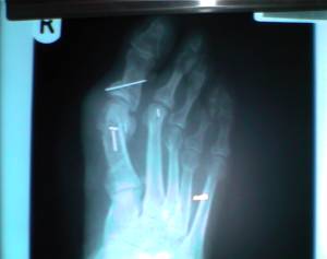
Conventional x-rays are the most common imaging technique for evaluating foot and ankle disorders. X-rays machines are readily available and exams are quick and relatively simple to perform. The denser an object is, the whiter it appears on an x-ray. Since bones are very dense, they are easily seen, but soft tissue abnormalities are not easily detected. X-rays excel at identifying fractures, arthritis, and bone tumours, but are not particularly useful in evaluating tendon and ligament injuries, masses, and infection. X-Ray showing surgical correction of a hallux valgus deformity. Also congenitally shortened bones of the forth and fifth metatarsal a condition known as brachymetatarsia. Please also note the pins and screws within this patient’s foot.
Conventional x-rays are the most common imaging technique for evaluating foot and ankle disorders. X-rays machines are readily available and exams are quick and relatively simple to perform. The denser an object is, the whiter it appears on an x-ray. Since bones are very dense, they are easily seen, but soft tissue abnormalities are not easily detected. X-rays excel at identifying fractures, arthritis, and bone tumours, but are not particularly useful in evaluating tendon and ligament injuries, masses, and infection. X-Ray showing surgical correction of a hallux valgus deformity. Also congenitally shortened bones of the forth and fifth metatarsal a condition known as brachymetatarsia. Please also note the pins and screws within this patient’s foot.
Nuclear Medicine
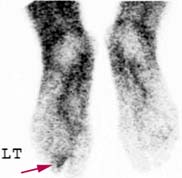
A tiny amount of radioactive material is injected through a vein and a special camera hooked up to a sensitive radiation detector is used to acquire images of the body. For foot and ankle imaging, the most common nuclear medicine study ordered is a "bone scan". The injected radiotracer is bound to a molecule that seeks out areas of high bone turnover. Increased radioactivity is detected at fractures, bone infections, and active arthritis. Bone scans are very sensitive tests and are able to detect bone abnormalities weeks before they are visible on conventional x-rays. A "triple-phase bone scan" obtains images at three different times during a four-hour period to allow differentiation between soft tissue infection (cellulitis) and bone infection (osteomyelitis). Since some patients who are suspected of having an infection may also have fracture or arthritis, this test may pose a diagnostic dilemma. It is therefore essential that x-rays are available when the test is being interpreted to assist in making an accurate diagnosis. Another nuclear medicine test which is used to evaluate for infection is called a "white blood cell" (WBC or Ceretec) scan. This test reveals abnormalities related to the presence of large numbers of white blood cells, and is therefore fairly specific in diagnosing infection. Nuclear medicine tests are commonly used to diagnose bone infection, but MRI is very popular because of its ability to provide superior spatial resolution and anatomic depiction. A Nuclear Medicine Bone scan note that both feet have been scanned. At the tip of the red arrow a marked darkened area can be seen. This is active bone infection within a diabetic patient.
A tiny amount of radioactive material is injected through a vein and a special camera hooked up to a sensitive radiation detector is used to acquire images of the body. For foot and ankle imaging, the most common nuclear medicine study ordered is a "bone scan". The injected radiotracer is bound to a molecule that seeks out areas of high bone turnover. Increased radioactivity is detected at fractures, bone infections, and active arthritis. Bone scans are very sensitive tests and are able to detect bone abnormalities weeks before they are visible on conventional x-rays. A "triple-phase bone scan" obtains images at three different times during a four-hour period to allow differentiation between soft tissue infection (cellulitis) and bone infection (osteomyelitis). Since some patients who are suspected of having an infection may also have fracture or arthritis, this test may pose a diagnostic dilemma. It is therefore essential that x-rays are available when the test is being interpreted to assist in making an accurate diagnosis. Another nuclear medicine test which is used to evaluate for infection is called a "white blood cell" (WBC or Ceretec) scan. This test reveals abnormalities related to the presence of large numbers of white blood cells, and is therefore fairly specific in diagnosing infection. Nuclear medicine tests are commonly used to diagnose bone infection, but MRI is very popular because of its ability to provide superior spatial resolution and anatomic depiction. A Nuclear Medicine Bone scan note that both feet have been scanned. At the tip of the red arrow a marked darkened area can be seen. This is active bone infection within a diabetic patient.
Magnetic Resonance Imaging (MRI)
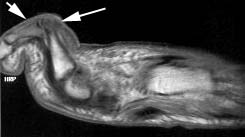
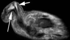
MRI has become the most robust imaging tool for diagnostic problem solving in the foot and ankle. MRI uses sophisticated computer technology coupled to powerful magnets to detect water and fat molecules, producing detailed, high resolution images of structures. MRI is a very sensitive test available for establishing whether an abnormality exists in the bone, soft tissues, tendons, or ligaments. MRI is capable of not only detecting an anatomic abnormality, but excels at characterising masses and evaluating the extent of soft tissue injury. Its ability to identify the type of tissue in a mass can indicate the likelihood of whether it is benign (not cancer) or aggressive (cancer, infection). Images are obtained in multiple cross sectional planes and different sequences are chosen to best characterize anatomy and abnormalities. Because of the strong magnetic field, patients with pacemakers and other implanted electronic devices are unable to have MRI. A T1W image, normal bone marrow, normal bone marrow in the toe is white and the abnormal marrow (arrows) is dark.This fat-suppressed T2W MRI image depicts normal bone marrow as dark and abnormal marrow as white (arrows). The MRI was able to assist the surgeon by demonstrating which part of the toe was infected and which part could be saved
MRI has become the most robust imaging tool for diagnostic problem solving in the foot and ankle. MRI uses sophisticated computer technology coupled to powerful magnets to detect water and fat molecules, producing detailed, high resolution images of structures. MRI is a very sensitive test available for establishing whether an abnormality exists in the bone, soft tissues, tendons, or ligaments. MRI is capable of not only detecting an anatomic abnormality, but excels at characterising masses and evaluating the extent of soft tissue injury. Its ability to identify the type of tissue in a mass can indicate the likelihood of whether it is benign (not cancer) or aggressive (cancer, infection). Images are obtained in multiple cross sectional planes and different sequences are chosen to best characterize anatomy and abnormalities. Because of the strong magnetic field, patients with pacemakers and other implanted electronic devices are unable to have MRI. A T1W image, normal bone marrow, normal bone marrow in the toe is white and the abnormal marrow (arrows) is dark.This fat-suppressed T2W MRI image depicts normal bone marrow as dark and abnormal marrow as white (arrows). The MRI was able to assist the surgeon by demonstrating which part of the toe was infected and which part could be saved
Computed Tomography (CT Scan)
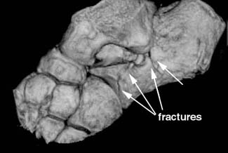
CT uses x-rays and a sophisticated computer to provide cross-sectional images. Where conventional x-rays superimpose overlying structures on top of one another, CT acquires thin (between 1 and 10mm) cross-sectional slices, allowing detailed evaluation of anatomy. For this reason, CT is the best exam for precisely determining the extent of a complicated fracture. Other uses for CT scans of the foot and ankle are to evaluate for fusion of joints (tarsal coalition), to quantitative the degree and location of arthritis at a joint, and for localization of a foreign body. While soft tissues are also visible, CT is not as capable at characterizing soft tissue abnormalities as MRI. Advanced computer post-processing allows CT images to be reconstructed in any plane, and can even create a three dimensional image, which is especially useful for surgical planning (FIGURE). Helical (or spiral) CT scanners are able to acquire images much more quickly than conventional CT scanners, and provide the best image reconstructions. Three-dimensional reformations generally require a helical (spiral) CT scanner and a dedicated computer workstation. 3D CT reformation of complicated calcaneus (heel) fracture shows involvement of multiple joints which were not apparent on regular X-rays. The images can be rotated in any direction and angle to allow optimal viewing of the fracture sites.
CT uses x-rays and a sophisticated computer to provide cross-sectional images. Where conventional x-rays superimpose overlying structures on top of one another, CT acquires thin (between 1 and 10mm) cross-sectional slices, allowing detailed evaluation of anatomy. For this reason, CT is the best exam for precisely determining the extent of a complicated fracture. Other uses for CT scans of the foot and ankle are to evaluate for fusion of joints (tarsal coalition), to quantitative the degree and location of arthritis at a joint, and for localization of a foreign body. While soft tissues are also visible, CT is not as capable at characterizing soft tissue abnormalities as MRI. Advanced computer post-processing allows CT images to be reconstructed in any plane, and can even create a three dimensional image, which is especially useful for surgical planning (FIGURE). Helical (or spiral) CT scanners are able to acquire images much more quickly than conventional CT scanners, and provide the best image reconstructions. Three-dimensional reformations generally require a helical (spiral) CT scanner and a dedicated computer workstation. 3D CT reformation of complicated calcaneus (heel) fracture shows involvement of multiple joints which were not apparent on regular X-rays. The images can be rotated in any direction and angle to allow optimal viewing of the fracture sites.
Ultrasound
Ultrasound is able to detect tendon abnormalities and masses, and has become a first line imaging modality for evaluation of sports medicine injuries The most common use for podiatric ultrasound is for detection of foreign bodies and to assist biopsy of masses usually neuromas which are benign nerve tumours which frequently occur within the metatarsal bones of the feet.
